The use of ionizing radiation in diagnostic imaging is a common practice worldwide. However, the imaging process itself carries a relative risk. Therefore, it is recommended to employ the lowest possible dose of ionizing radiation, especially in computed tomography (CT) imaging, where a series of X-ray scans are utilized to reconstruct body tissue sections.
A popular method for radiation dose reduction in CT imaging is known as the quarter-dose technique, which reduces the X-ray dose but can cause noise and loss of image sharpness. Since CT image reconstruction from directional X-rays is a nonlinear process, it is analytically difficult to correct the effect of dose reduction on image quality. Recent and popular deep learning approaches provide an intriguing possibility of low-dose CT image enhancement.
Some recent works propose combinations of multiple deep learning and classical methods for this purpose, which over-complicate the process. However, it is observed here that the straight utilization of the well-known U-NET provides very successful results for correcting low-dose artifacts.
Blind tests conducted with practicing radiologists demonstrate that U-NET-enhanced quarter-dose CT images not only show significant visual improvement compared to their low-dose counterparts but are also diagnostically superior, even when juxtaposed with full-dose CT images.
Methodology
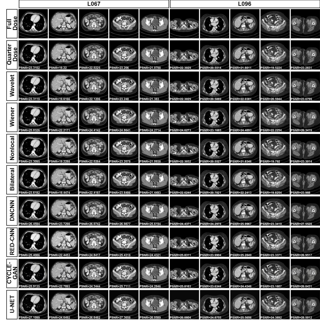
In this study, the quarter dose images were applied to four classical and four DL-based image enhancement methods and a comprehensive comparative evaluation was conducted based on their performances. The classical image processing-based enhancement methods were Wavelet denoising, Wiener filtering, Non-local means filtering, and Bilateral filtering, while RED-CNN, CycleGAN, DnCNN and U-NET were utilized as DL-based algorithms. Despite their typically lower performance compared to DL-based methods, these classical image processing techniques were incorporated into the study to provide a foundational baseline for comparison.
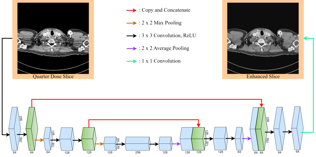
Among the DL-based methods, the U-NET model emerged as the preferable (hence the proposed) method due to its visibly superior enhancement performance, as well as its simplicity and ease of implementation using off-the-shelf software. This autoencoder-type DNN, initially designed for biomedical image segmentation, is comprised of a contracting (input-side) and an expansive (output-side) path, interconnected with auxiliary connections. The contracting path of U-NET consists of repeated 3×3 convolution layers, followed by a ReLU activation layer and a 2×2 max pooling layer with a stride of two. At the end of this path, features are collated into a single feature via a 1×1 convolution layer. Contrarily, the expansive path employs convolutional and average pooling layers to revert the image to its original size. A unique characteristic of U-NET is the transfer of certain features from the contracting to the expansive path, allowing for enhanced localization. An illustration of our utilized U-NET structure can be found in Figure 1, which displays both the low-dose input and enhanced output images, standardized at a resolution of 256×256 pixels.
For a fair comparison, all DL-based architectures were trained with the same regimen. They were trained for 100 epochs from scratch with training data comprising 5926 image pairs extracted from the Low Dose CT Grand Challenge dataset. Experimental ResultsIn this dataset, chest CT images were obtained from ten patients. Our study specifically targeted those images exhibiting a slice thickness of 1 mm.
Experimental Results
The experiments were conducted on a hardware equipped with a Windows 10 operating system, four 32 GB Nvidia GeForce GTX 1080 GPUs, 32 GBs of RAM, and a 6-core 5th generation Intel Core i7 CPU with a 3.5 GHz clock speed.
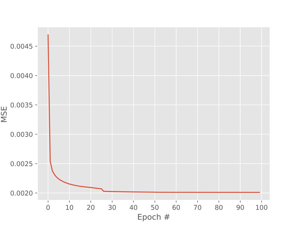
In order to make a fair assessment, the DNNs (except the DnCNN, which is already a pre-trained network for general de-nosing purposes) were trained for 100 epochs with an optimization criterion of mean-squared error (MSE) as the fidelity measure between the enhancement and the full-dose images. Figure 2 illustrates the evolution of the MSE values for 100 training epochs in U-NET. In this figure, the MSE begins at 0.0045 and converges to approximately 0.0020 after 30 epochs.
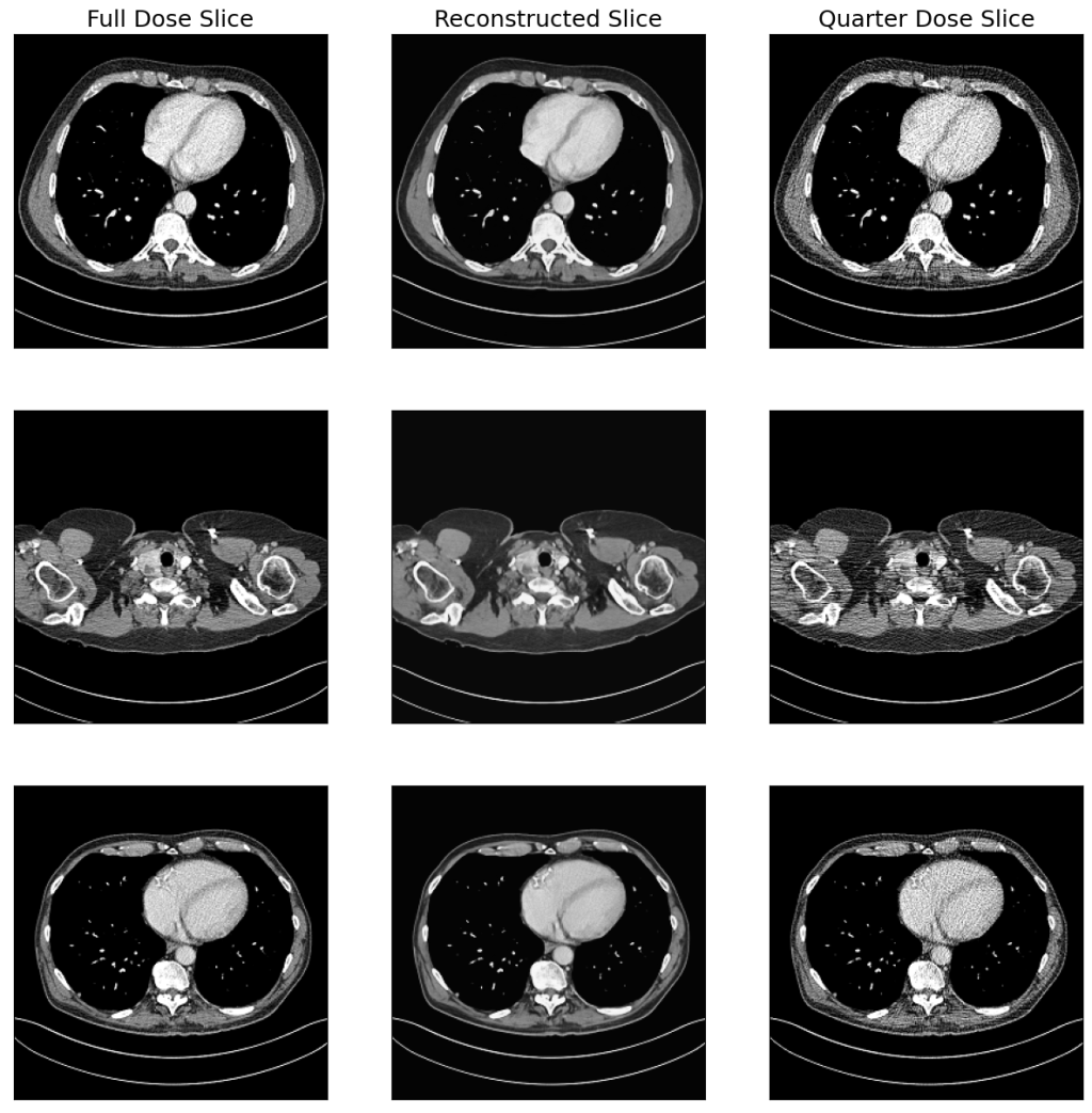
In the test phase, 50 exclusive quarter-dose and full-dose CT test images were used. The quarter-dose versions of the CT test images were fed to the proposed U-NET, as well as three more state-of-the-art DL-based algorithms and four classical image enhancement methods. The resultant images were saved for quantitative, qualitative, and comparative analysis. Figure 3 show full-dose, quarter-dose and enhanced versions of the CT test scans in columns.
Want to read more? Here is the link https://arxiv.org/abs/2310.20265


Leave a comment